Diagnostics

Eye Testing in Abu Dhabi
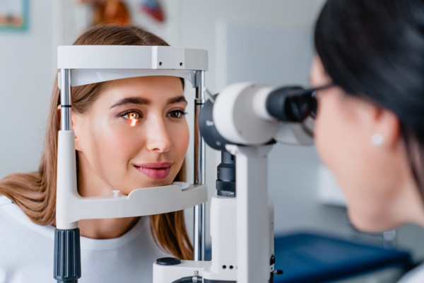
Dr. Madhava Rao, the best eye doctor in Abu Dhabi offers state-of-the-art diagnostic tools and unparalleled expertise with comprehensive eye care services tailored to focus on the unique needs of each patient for eye testing in Abu Dhabi.
From advanced retinal imaging systems to precision corneal topography and comprehensive visual field testing, these diagnostic tools enable us to detect and monitor various eye conditions with unparalleled precision. Get your diagnostics done by well-known ophthalmologist Abu Dhabi practicing in Burjeel hospital.
Wide Field Retinal Imaging
Retinal imaging is essential in ophthalmic practice, providing two-dimensional images of the complex three-dimensional retinal tissue. These techniques are invaluable for diagnosing, planning, monitoring and managing various eye and systemic conditions, including retinal detachment surgery in Abu Dhabi, diabetic retinopathy, age-related macular degeneration, retinal vein occlusion, glaucoma, other retinal dieases.
Optical Coherence Tomography
Optical coherence tomography (OCT) and optical coherence tomography angiography (OCTA) are non-invasive imaging methods utilizing light waves to capture retina cross-sections. OCT enables our ophthalmologist in Abu Dhabi to precise visualization and measurement of retinal layers, aiding in diagnosing and treating conditions such as diabetic macular edema (DME), age-related macular degeneration (AMD), various macular diseases, and glaucoma,
OCTA-OCT Angiography
Optical coherence tomography angiography (OCT-A) is a non-invasive method for imaging the retinal and choroidal microvasculature. It uses laser light reflectance to visualize moving red blood cells, providing detailed vessel images without injecting the colored dyes in vein.
Multicolor Imaging
Multicolor Imaging utilizes various light wavelengths to generate vivid colour images of the retina. This method improves the clarity of retinal structures and abnormalities, providing clinicians with a comprehensive view to evaluate different eye conditions.
Intravenous Fluorescein Angiography (IVFA/FFA)
Our ophthalmologist in Abu Dhabi uses Fluorescein Angiography (FA) for capturing retina images using a specialized camera, aiding in the detailed examination of blood vessels and ocular structures. It’s crucial for monitoring disease progression and identifying treatment sites.
Intravenous Indocyanine Green Angiography (ICGA)
ICGA uses indocyanine green dye to observe deeper ocular structures, primarily the choroid, aiding in the identification of conditions like choroidal neovascularization. This method offers supplementary insights beyond those acquired from fluorescein angiography.

Ultrasonography B Scan
B scan, or Bright Scan ultrasonography, is employed when viewing the back of the eye is challenging. It utilizes sound waves to create 2D images of ocular structures, bypassing dense pathology and can help diagnose ocular inflammation by visualizing the orbit and eye muscles.
Optical Biometry
Optical biometry is a precise and non-invasive automated technique used to measure the eye’s anatomical features. These measurements are crucial in determining the appropriate power of an intraocular lens (IOL) before cataract surgery.
Corneal Topography
Corneal topography utilizes a unique photographic method to chart the contours of the transparent front surface of the eye, known as the cornea. This process resembles creating a 3D representation akin to mapping out the peaks and troughs of terrain.
Specular Microscope
Specular microscopy is a method used to examine the density and structure of corneal endothelial cells. It plays a crucial role in assessing the health and performance of the cornea, aiding healthcare providers in monitoring conditions that impact the deepest layer of the cornea.
Pachymetry
Pachymetry is an eye examination that determines the thickness of the cornea, the transparent layer at the eye’s front. Typically, the central cornea measures about 500 to 600 microns, while the peripheral region ranges between 600 and 800 microns.
Humphrey's Visual Field
Humphrey’s Visual Field test examines the entire range of a person’s vision, identifying any areas of vision loss or abnormalities. This test is frequently employed to assess conditions like glaucoma and neuro-ophthalmic disorders, helping to detect and track these conditions early on.
Electrophysiological Studies
Electrophysiology serves as a diagnostic tool and monitors the advancement of vision-related disorders, much like how electrocardiograms (ECGs) track heart disease progression. Electrical signals are generated by the retina, optic pathways in the brain, and visual cortex. These signals are directly recorded from the eye or extracted via computer from brain electrical signals recorded from the scalp.
Eye Testing in Abu Dhabi | Best Eye Doctor in Abu Dhabi - Dr Madhava Rao
Want to know more about these vision testing diagnostic tools? Consult Dr. Madhava Rao – the best eye doctor in Abu Dhabi!
Fellowships & Memberships


Accolades - Your Eyes are in Award-winning Hands

Ophthalmologist Education Award
A testament to Dr. Rao's commitment to advancing ophthalmic education and sharing knowledge with the next generation of eye care professionals.

International Ophthalmology Scholar Award
Recognizing his significant contributions to the global ophthalmology community, this award highlights Dr. Rao's role in enhancing the understanding and treatment of eye diseases worldwide.

