Retinal Vascular Occlusion: Symptoms, Causes, Treatment & When to See a Specialist
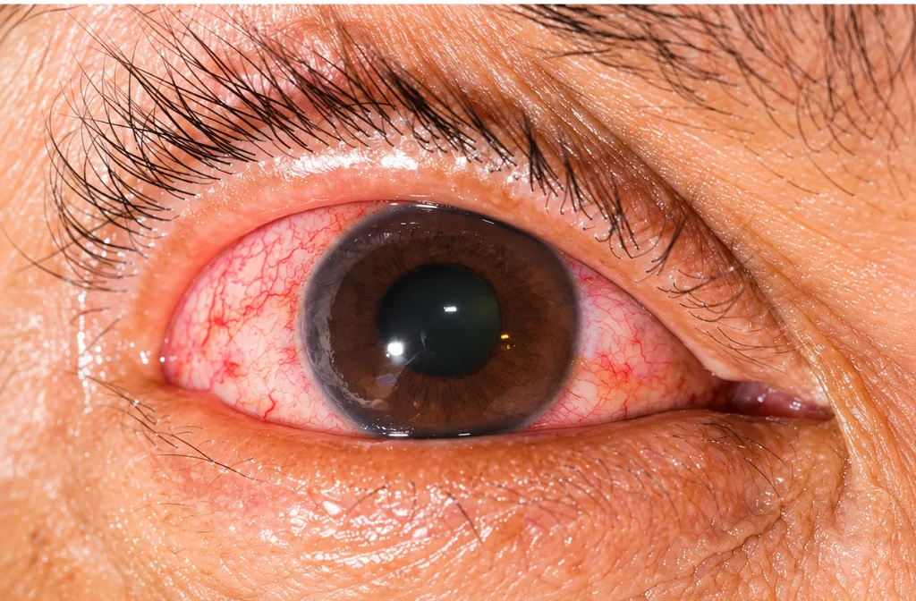
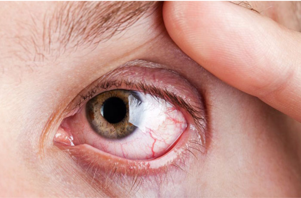
Retinal vascular occlusion occurs when a blood vessel supplying or draining the retina becomes blocked. Because the retina depends on a continuous blood supply, a blockage can affect vision and may lead to retinal or macular complications.
What Is Retinal Vascular Occlusion?
The retina is the light-sensitive tissue at the back of the eye. Small arteries deliver oxygen-rich blood to it, while veins carry blood away. When one of these vessels becomes blocked, retinal circulation is disrupted.
The effect on vision depends on the vessel involved, the location of the blockage and whether complications such as macular edema, retinal ischemia or abnormal blood-vessel growth develop.
Types of Retinal Vascular Occlusion
Retinal Vein Occlusion
A retinal vein occlusion (RVO) occurs when a retinal vein is blocked. It may involve the main retinal vein or one of its branches.
- Central retinal vein occlusion (CRVO): the main retinal vein is affected.
- Branch retinal vein occlusion (BRVO): a smaller branch of the retinal vein is blocked.
A vein blockage can cause blood and fluid to leak into the retina. When the macula becomes swollen, known as macular edema, central vision may become blurred or reduced.
Retinal Artery Occlusion
A retinal artery occlusion (RAO) occurs when blood flow through a retinal artery is blocked. Central retinal artery occlusion can cause sudden, significant loss of vision in one eye, while a branch retinal artery occlusion may produce a missing area in part of the visual field.
This type of sudden vision loss should be treated as an emergency because retinal artery occlusion can be associated with stroke and other vascular disease.
What Causes Retinal Vascular Occlusion?
The risk factors vary according to the type of occlusion. Conditions that can contribute to retinal vascular disease include:
- High blood pressure
- Diabetes
- High cholesterol and other vascular risk factors
- Cardiovascular disease
- Glaucoma, particularly in relation to retinal vein occlusion
- Smoking
- Obesity and other metabolic risk factors
Because retinal artery occlusion can be linked to systemic vascular disease, an eye assessment may be followed by medical evaluation of cardiovascular or stroke risk.
What Are the Symptoms?
Symptoms depend on the type and severity of the blockage. Possible symptoms include:
- Blurred or reduced vision in one eye
- Sudden, painless loss of vision
- A missing or dark area in the visual field
- Difficulty seeing fine details
- New or increased floaters in some cases
Retinal vein occlusion may cause blurred vision as retinal or macular swelling develops. Retinal artery occlusion more typically causes sudden, painless visual loss or a sudden visual-field defect.
Sudden Vision Loss Should Never Be Ignored
If vision suddenly becomes significantly blurred or is lost in one eye, do not wait to see whether it improves on its own. Acute retinal artery occlusion needs immediate medical evaluation and may require assessment at a stroke centre or other emergency facility.
How Is Retinal Vascular Occlusion Diagnosed?
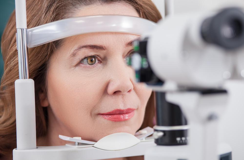
A retina specialist will review the symptoms and medical history and examine the retina. Depending on the findings, testing may include:
Dilated Retinal Examination
Dilating the pupils allows the ophthalmologist to examine the retina and retinal blood vessels for bleeding, swelling, ischemia and abnormal blood-vessel growth.
Optical Coherence Tomography (OCT)
OCT provides detailed cross-sectional images of the retina. It is particularly useful for detecting and monitoring macular edema and other structural changes.
Fluorescein Angiography
In selected cases, fluorescein angiography can help assess retinal blood flow, leakage and areas of reduced perfusion.
The tests required depend on the type of occlusion, symptoms and examination findings.
How Is Retinal Vascular Occlusion Treated?
There is no single treatment for every case. Management depends on whether the blockage involves a vein or artery and whether complications such as macular edema or neovascularisation are present.
Treatment of Retinal Vein Occlusion
When retinal vein occlusion causes macular edema, anti-VEGF injections are commonly used to reduce retinal swelling and protect or improve vision. Corticosteroid treatment may be considered in selected patients. Laser treatment may also have a role in particular complications.
Follow-up is important because retinal vein occlusion can change over time. The ophthalmologist may monitor for macular edema, retinal ischemia and abnormal blood-vessel growth.
Treatment of Retinal Artery Occlusion
Retinal artery occlusion requires a different approach. The priority is urgent systemic and vascular evaluation rather than relying on routine eye treatment alone. There is no established treatment that reliably reverses the retinal damage once an acute artery occlusion has occurred, so rapid assessment for associated vascular disease is critical.
Can Retinal Vascular Occlusion Be Prevented?
Not every case can be prevented, but controlling vascular and metabolic risk factors is an important part of reducing risk.
- Monitor blood pressure and blood sugar.
- Manage cholesterol and cardiovascular risk factors.
- Avoid smoking.
- Attend regular eye examinations when you have known eye or systemic risk factors.
When Should You See a Retina Specialist?
Arrange an eye examination for unexplained or persistent changes in vision. Sudden or significant vision loss in one eye is different: it requires urgent medical attention and should not be treated as a routine appointment.
Retinal Vascular Occlusion Treatment in Abu Dhabi
At RetinaCare Abu Dhabi, patients can receive specialist retinal evaluation for conditions affecting the retina and vision. A retina specialist can assess the condition, recommend appropriate diagnostic testing and discuss treatment and follow-up according to the individual findings.
If you experience sudden vision loss or a major change in vision, seek urgent medical attention rather than waiting for a routine consultation.
FAQs
Q1. What is retinal vascular occlusion?
It is a blockage in a blood vessel supplying or draining the retina. It may affect a retinal artery or vein and can cause changes in vision.
Q2. What are the symptoms of retinal vascular occlusion?
Symptoms may include blurred or reduced vision, sudden vision loss, visual-field defects and, in some cases, new or increased floaters.
Q3. Is retinal vascular occlusion serious?
Yes. The seriousness depends on the type and severity. Retinal artery occlusion can cause sudden severe vision loss and requires urgent evaluation. Retinal vein occlusion can lead to macular edema and other retinal complications.
Q4. How is retinal vein occlusion treated?
Treatment depends on the retinal findings. Anti-VEGF injections are commonly used when macular edema is present; other treatments may be considered for selected complications.
Q5. Is retinal artery occlusion an emergency?
Yes. Sudden symptomatic retinal artery occlusion is an ophthalmic emergency and requires immediate medical evaluation, including assessment for associated vascular or stroke risk.
Conclusion
Retinal vascular occlusion can affect vision when a retinal artery or vein becomes blocked. Retinal vein occlusion may cause macular swelling, while retinal artery occlusion can produce sudden vision loss and requires urgent assessment.
Early recognition and appropriate retinal evaluation are important. If you notice a sudden or unexplained change in vision, do not delay seeking professional eye care.